
Coccidial monitoring of chickens and turkeys is done by routine intestinal lesion scoring, typically at 28 days of age.
The most widely used system for coccidial monitoring is the Johnson and Reid Scoring System. Using this system, lesion scores, noted from one to four (zero for a normal appearance of the intestine), are assigned for each of the five coccidial species likely to induce lesions in chickens. A similar system was developed for turkeys by ANSES, France.
Visible lesions in the gut mucosa typically appear in late schizogony or during gamogony, and their severity remains constant for a few days. The lesions are caused by the release of the merozoites and eventually the unsporulated oocysts. (A lesion score of three does not result from a lesion score of two the previous day, and will not evolve into a different severity the following day in the same bird). However, as the epithelium regenerates, the lesions slowly disappear making accurate scoring more difficult.
Lesion Scores
Lesion scores are useful for:
- Assessing the severity of coccidial development
- Identifying the coccidial species involved, and
- Establishing a link between the parasite manifestation observed on the farm and in production
Chicken Lesion Scores
Photos credits: Chicken Lesion Scores / Jean-Michel Répérant
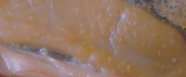
E. acervulina
Score 1
Scattered white plaques on the mucosal surface, mainly in the duodenum. Fewer than 5 lesions per cm2.
Score 2
More numerous white plaques in the duodenum and/or jejunum, but not coalescent. Normal color. Criterion: Score 2 when white plaques number more than five in 1 cm of intestinal length (whatever the intestinal portion).
Score 3
White plaques are numerous enough to coalesce at some sites AND mucosal discoloration is observed. Contents are watery. The mucosa may be covered with a whitish coating, but the score remains 3 as long as white plaques appear on the epithelium under this coating.
Score 4
Discrete white plaques are no longer visible. Mucosal discoloration is extreme and the mucosa may be covered with a whitish coating laden with oocysts (microscopic confirmation).
The coating may have disappeared, making the lesion arduous to score. Compare with other intestines before giving a final score.
E. brunetti
Score 1
A few petechiae on the serosal surface, mostly at the ileal level, but which can be found in other portions of the intestine.
Score 2
More numerous petechiae on the serosal surface, mostly at the ileal level, watery contents and petechiae on the mucosal side sometimes extend down to the large intestine
Score 3
Petechiae on the serosal surface or ileum light red in color, numerous small petechiae on the mucosal surface, watery contents with abundant orange- or bloodtinged mucous, thick cecal contents with traces of fibrin or blood. Ballooning of the intestine.
Score 4
roughened mucous membrane, numerous small petechiae on the mucosal surface, watery and mucoid contents, hemorrhages or epithelial necrosis, cecal contents mixed with blood or fibrin. Ballooning of the intestine.
E. maxima
 Score 1
A few petechiae on the serosal surface around Meckel’s diverticulum, or in other areas of the intestine.
Score 2
More numerous petechiae on the serosal surface, small petechiae on the mucosal side, watery contents, orange mucus. Thickened intestinal wall.
Score 3
Ballooning of the small intestine and thickening of the intestinal wall. Abundant orange mucus, traces of blood in mucus.
Score 4
significant ballooning, blood and blood clots scattered in orange mucus, watery contents, putrid odor.
E. necatrix
Score 1
Scattered white spots and petechiae on the serosal surface around Meckel’s diverticulum.
Score 2
Numerous white plaques and petechiae on the serosal surface, slight ballooning. Somewhat increased mucus secretion.
Score 3
Very numerous white plaques and petechiae on the serosal surface. Increased ballooning of the ileum. Extensive hemorrhages and abundant mucus in the lumen.
Score 4
Extensive ballooning and purple color of the intestine. Contents mixed with very abundant mucus, red or brown in color, with massive hemorrhages.
E. tenella
Score 1
Scattered petechiae on the cecal serosal and mucosal surfaces. Little blood in the ceca. Thick cecal contents.
Score 2
Petechiae on the cecal serosal and mucosal surfaces. Thick cecal contents containing blood or fibrin. Thickened wall, but presence of grooves.
Score 3
Cecal wall greatly thickened, grooves no longer visible. Absence of cecal contents replaced by blood or fibrin.
Score 4
Distended and club-shaped ceca. The lumen of the distended ceca is all filled with blood (schizogony) or blood clots.
Turkey Lesion Scores
Photos credits: Turkey Lesion Scores / Jean-Michel Répérant
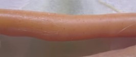
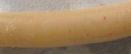
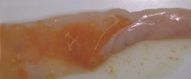
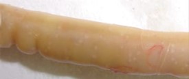
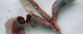
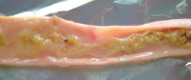
E. adenoeides
Score 1
Caeca appear normal. Caecal contents normal, that surrounds some "grains" of caseous material. No lesions in the end of the intestine
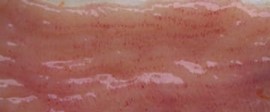
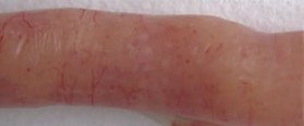
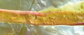
Score 2
Caeca has a clearer appearance. Fragments of caseous material in content that is more liquid. Some petechiae on the furrows of the caecal mucosa. No lesions in the end of the intestine.
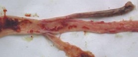
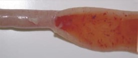
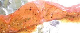
Score 3
Caeca is much paler and less flexible. Contents replaced by a lot of caseous material. Caseous material more or less compact, immersed in a clear liquid. Blood may be mixed in the liquid. The mucosal grooves are visible but covered with petechiae. Numerous petechiae in the terminal part of the intestine.
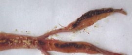
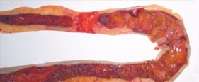
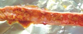
Score 4
Caeca is whitish. They are filled with caseous material. Mucosa is very fine and completely leveled. Blood can be mixed with the caseous material. The terminal part of the intestine is bloody.
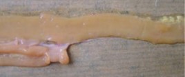
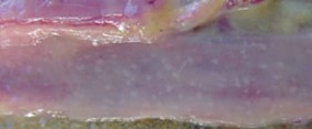
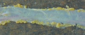
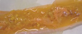
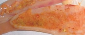
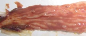
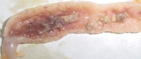
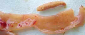
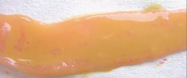
E. meleagrimitis
Score 1
Some filaments in the duodenal loop. Content in the jejunum is food. Normal mucosa.
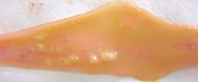
Score 2
Little or no food content. Normal mucosa in duodenum, but finer in the jejunum. Presence of many filaments which can form fine pseudo-membranes, but not orange.
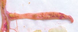
Score 3
Little or no food content. Mucosa is leveled, pseudo-membranes is orange, thick strands in descending duodenum. No blood.
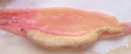
Score 4
Mucosa very leveled, no food content. A lot of pseudo-membranes observed. Duodenal loop obstructed. Petechiae visible. Liquid colorless but presence of blood or melena.
![Coccidiosis extrait[2259]-1](https://poultrycontent.ceva.com/hs-fs/hubfs/Coccidiosis%20extrait%5B2259%5D-1.png?width=951&height=862&name=Coccidiosis%20extrait%5B2259%5D-1.png)
![Coccidiosis extrait[2259]-1](https://4368135.fs1.hubspotusercontent-na1.net/hubfs/4368135/Coccidiosis%20extrait%5B2259%5D-1.png)