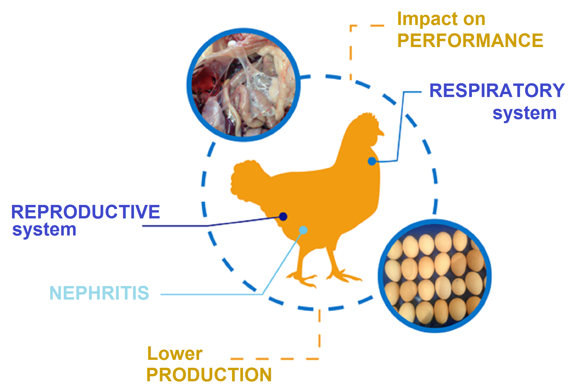
One of the most important steps for the control of a disease in poultry production is to ensure accurate diagnosis. Infectious Bronchitis (IB) should be distinguished from other clinical diseases causing respiratory signs and lesions, Nephrosonephritis and impact in egg production and quality.
Some of the other respiratory diseases commonly found in poultry can include Newcastle disease, caused by lentogenic respiratory strains, low pathogenicity avian influenza, infectious laryngotracheitis, swollen head syndrome, Mycoplasma gallisepticum infection.
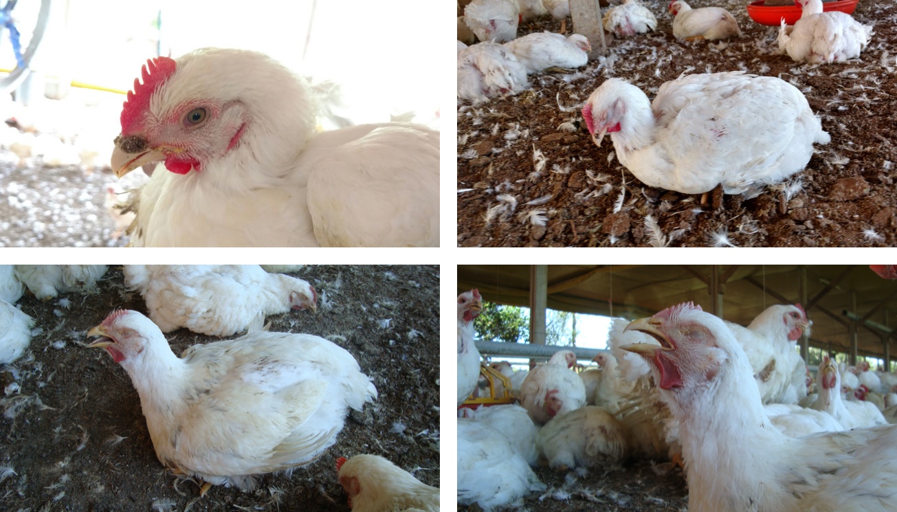 Images of signs of IB infection, by Chacón.
To detect the IB infection, and differentiate it from other diseases, clinical teams should look at the bird’s history, clinical signs and lesions. To confirm the initial diagnosis, laboratory tests will assist.
Serological test will demonstrate the response of the chickens’ immune system to the virus by the increase of titers in IB ELISA tests. The natural infections will generate a high level of titers.
It’s important to note that a differentiation should be done, as live attenuated and inactivated vaccines aim to generate humoral protection, by increasing antibodies, that can be observed by titers in serological tests. Different vaccines will produce different levels of titers according to the test used.
If possible the diagnosis can identify the virus’ serotype or genotype, in light of the extremely high rate of antigenic variation among IBV strains and the different serotype-specific vaccines (Cavanagh & gelb Jr., 2008). When taking samples for IBD identification, these should be collected as soon as possible and frozen for delivery to the lab.
For live birds, swabs should be taken from the upper respiratory tract. From carcasses, tissue samples from the trachea and the lungs should be used. In birds exhibiting nephritis or egg production problems, samples should be collected from the kidneys and the oviduct as well.
If a histopathological exam is conducted, performance can be observed due to the damage caused by the virus replication. As well as impacting the ciliostasis score, it means lower mucosa cilios movements beats.
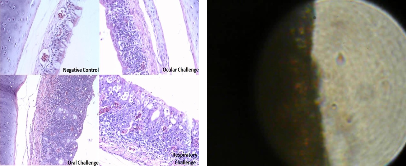
In the left image of histopathological examination, and in the right ciliostasis mucosa exam.
Another technique that can be used is the reverse transcription-polymerase chain reaction (RT-PCR), using respiratory or cloacal swabs, fresh carcass kidney or cecal tonsils.
For genotyping, the characterization of strains will be based on part of the genome. Due to the high variability between IBV strains, using a more stable part of the genome avoids the need to produce high demand for primers, as it will be able to detect all different strains.
One of the possible approaches that can be used for the RT-PCR for the S1 gene is to look for expected and known strains. This way the confirmation of the diagnosis will be facilitated in most cases.
When differentiating from other diseases, not just respiratory virus should be the focus. Nephrosonephritis caused by nephropathogenic IBV infection are detected in birds older than 4–6 weeks of age. They should be differentiated from infectious bursal disease (Gumboro), mycotoxicoses (ochratoxin a) etc.
When differentiating from other diseases, not just respiratory virus should be the focus. Nephrosonephritis caused by nephropathogenic IBV infection is detected in birds older than 4–6 weeks of age. They should be differentiated from infectious bursal disease (Gumboro), mycotoxicoses (ochratoxin a) etc.